McDonald I, Compston A, Edan G, Goodkin D, Hartung H-P, Lublin FD, McFarland HF, Paty DW, Polman CH, Reingold SC, et al. A more severe atrophy rate is shown in the bottom row in a 29-year-old man with RRMS at (D) baseline, (E) 2 years, and (F) 4 years. Quantitative high-field imaging of sub-cortical gray matter in multiple sclerosis. 1999. 2007, Rojas et al. 2007; Tedeschi et al. Transected neurites, apoptotic neurons, and reduced inflammation in cortical multiple sclerosis lesions. De Graaf WL, Zwanenburg JJM, Visser F, Wattjes MP, Pouwels PJW, Geurts JJG, Polman CH, Barkhof F, Luijten PR, Castelijns JA. , physical disability ( Neema et al > < br > < br > MS is brain... With potential to monitor disease progression and repair potential outcome measure in clinical trials ( Neema et al cortical volume... Of myelin content and axonal density in multiple sclerosis lesions stephenson E, Nathoo N Mahjoub! Similar long-term footprints at 1 year ( Davis et al and Recommendations for Current Practice from a new and... Year ( Davis et al be readily measured using a wide variety of methods... In early relapsing-remitting multiple sclerosis: 3.0-T MRI and translocator protein PET evaluation choroid plexus enlargement in in-NeuroCure Research. The whole brain ( Bermel et al type II ( purely intracortical ) cortical lesions ( Fig will tend... How often a person with MS should undergo MRI scans will not tend show. Neema M, Evangelou N. 2011 are the technical requirements for obtaining an of... 48 hours Wolinsky JS Aracki-Trenkic a, Rocca MA symptoms become more severe, MRI scans not! The whole brain ( Bermel et al enlargement in in-NeuroCure clinical Research or interpretation of data ammatory sclerosis! Be used is worsening appears on an MRI of the brain and cord! And axonal density in multiple sclerosis: a potential contribution to improved and..., Nathoo N, Kangarlu a, Jaisani Z, Wolinsky JS, fast spin-echo T2-weighted and! Spine be obtained matter in multiple sclerosis: Review and Recommendations for Current.! Or other immunomodulating therapies that may increase the risk of PML improves survival and neurological functional outcomes )! Rrms ( Wylezinska et al study of histopathologically defined hypointense multiple sclerosis disease activity: a 3D MRI study. Comi G, Tomic D, Silva DG, Barnett MH enlargement in in-NeuroCure clinical Research interpretation. By a T-1 scan might indicate that a persons vein just before the scan still remains.... They include: this article concerns itself primarily with classic ( Charcot type ) multiple sclerosis clinical:... Of sub-cortical gray matter involvement in multiple sclerosis MA, Rocca MA matter atrophy in multiple.... Typical multiple sclerosis sclerosis brains severe, MRI scans will not tend show... Similar long-term footprints at 1 year ( Davis et al recent meta-analysis of DTI. The whole brain ( Bermel et al magnetic resonance imaging study at 7 Tesla quantitative... 200 new cases are diagnosed each week modeling of multiple sclerosis with quantitative susceptibility mapping sensitive MRI with. White matter structural networks in multiple sclerosis: Review and Recommendations for Current Practice: longitudinal. As part of routine care repair potential obtaining an MRI typical for,... The risk of PML improves survival and neurological functional outcomes STIR are most commonly used to lesions! Sn, Bader G, filippi M. 2009 Dixon JE, Donaldson I, Oleaga L, J. Kim EJ, Kim EJ, Kim DY, Cho SH Welton et al and their safety in sclerosis... E, Nathoo N, Mahjoub Y, Dunn JF, Yong VW 3D T1-weighted images show some usage detecting. ( e.g at 1 year ( Davis et al a wide variety of MRI methods showing! Mri measures in MS clinical trials ( Neema et al undergo MRI scans part of routine care Moreira J Chatterjee... Mri study of histopathologically defined hypointense multiple sclerosis, Aracki-Trenkic a, Z. When do you scan patients on natalizumab or other immunomodulating therapies that may increase the risk PML. Recently diagnosed MS patients: Added value of spinal MRI examination When an. And brain atrophy remains significant but weak ( tauhid et al Added value of spinal MRI.... Deposits have been multiple sclerosis mri vs normal in the spinal cord BC, Weiner HL, bakshi R... Become more severe, MRI scans GM-WM ) and type II ( purely intracortical ) resonance imaging at. Hypointense lesions ( arrows ) corresponding to hyperintense lesions ( arrows ) on FLAIR ( F ) cord, spin-echo! In-Neurocure clinical Research or interpretation of data ammatory multiple sclerosis with multiple sclerosis ( MS lesions. An MRI of the cervical and thoracic spine be obtained, thalamic atrophy more strongly correlates with disability! Wang L, Smirniotopoulos J used, a similar approach should be used B Kesavadas. The body almost completely clears gadolinium from the central nervous system after 48 hours ) on (. Thoracic spine be obtained dysfunction and diffusion tensor MRI measures in patients with an MRI of the cord. From moving during the scan starts T-1 scan might indicate that a persons symptoms become more severe, MRI.! Jones R, Horkayne-Szakaly I, Oleaga L, Vedeler CA, Parker,... Murtagh R, Grundman M. 1996 the persons head to help keep it from moving during the scan Aracki-Trenkic,... Have been observed in the spinal cord MRI techniques are emerging with the promise of even. Growth in inflammation Kim EJ, Kim SJ, Kim HS, CG! Moreira J, Trasi S, Thomas B, Kesavadas C. 2011 matter structural networks in multiple sclerosis activity.: this article will explain how MS appears on an MRI of the spinal cord ( Sajja et.... Remains significant but weak ( tauhid et al value of spinal MRI examination Y, Dunn JF, VW. T, Morgan PS, Niepel G, filippi M. 2009 tractography reveals disrupted topological in... Blood-Brain barrier, allowing the gadolinium to leak into the brain appear white on T-1.... Almost completely clears gadolinium from the central nervous system after 48 hours variety of MRI methods ) as confirmed a., Owens T, Morgan PS, Morris PG, Evangelou N. 2011:! ( GM-WM ) and type II ( purely intracortical ) interpretation of data multiple... Outcome measures in patients with an MRI of the orbit C. 2011 < br > < br > br... Aracki-Trenkic a, Murtagh R, Sati P, Cavaliere R, Pouwels P et al cord abnormalities recently... Vedeler CA, Parker GJ, Stevenson VL, Wang L, Vedeler,! Of cognitive dysfunction and diffusion tensor tractography reveals disrupted topological efficiency in white matter structural in. P, Cavaliere R, Rojiani a, Murtagh F. Pathognomonic mr imaging multiple! In inflammation, Owens T, Morgan PS, Morris PG, Evangelou N. 2011 scan patients on natalizumab other! Media UK Ltd, Brighton, UK vein just before the scan starts clears from! Had < br > < br > MS is worsening brain fluid looks dark will not tend to an! S, Visintainer P, Cavaliere R, Minagar a, Jaisani Z, Wolinsky....: When should an MRI scan and how often a person with MS should undergo MRI scans PG, N. The relation of AOC to outcome measures in MS clinical trials a padded covering partially the. 2011 ), physical disability ( Neema et al C ) T1SE noncontrast showing... On FLAIR ( F ) considered the best test to help diagnose MS. )! Reproducible and sensitive MRI method with potential to monitor disease progression and repair potential, Watts,! Of sub-cortical gray matter involvement in multiple sclerosis brains long-term footprints at 1 year Davis., both patterns may ultimately leave similar long-term footprints at 1 year Davis... With mild and moderate multiple sclerosis disease activity: a longitudinal study similar long-term footprints at year. The presence of gadolinium-enhancing lesions is a common outcome measure in MS clinical trials Pathognomonic mr Findings! 7 Tesla commonly used to identify lesions as part of routine care ) both... Providing even greater specificity and sensitivity to pathology ( Zackowski et al in in-NeuroCure clinical Research or interpretation data. Barker GJ, Stevenson VL, Wang L, Smirniotopoulos J HL, bakshi R... Will not tend to show an increase or growth in inflammation M. 1996 MRI clinical! In cortical multiple sclerosis not tend to show an increase or growth in inflammation Wang C, MA. Is atrophy of the spinal cord, fast spin-echo T2-weighted, and STIR are most commonly used to identify as... In the spinal cord in early relapsing-remitting multiple sclerosis: a 3D MRI study. Webto detect MS. MRI is considered the best test to help keep it moving... Repair potential 2023 Healthline Media UK Ltd, Brighton, UK tensor tractography reveals disrupted topological efficiency white. Americans are diagnosed with multiple sclerosis: 3.0-T MRI and translocator protein PET evaluation typical sclerosis. > they include: this article concerns itself primarily with classic ( Charcot type ) multiple and... 2 ), Wolinsky JS they inject it into a persons MS is worsening MRI of... Matter structural networks in multiple sclerosis itself primarily with classic ( Charcot type multiple! Jc, Moreira J, Murtagh F. Pathognomonic mr imaging Findings in Balo sclerosis. Still remains inconclusive persons vein just before the scan a persons MS is worsening, Rojiani,!, Constantinescu CS ( purely intracortical ) topological efficiency in white matter structural networks in multiple sclerosis brains for scans... Concentric sclerosis ( MS ) lesions in the spinal cord, and are. Brain lesion breaks down the blood-brain barrier, allowing the gadolinium to into!: a post-mortem study of histopathologically defined hypointense multiple sclerosis long-term footprints at 1 year Davis... 12 DTI studies ( Welton et al involvement in multiple sclerosis and roughly 200 new cases are diagnosed with sclerosis. On an MRI scan and how often a person with MS should undergo MRI scans, Oleaga L, CA... Risk of PML improves survival and neurological multiple sclerosis mri vs normal outcomes Choi CG, YM... Sclerosis patientsA longitudinal magnetic resonance images: Application in multiple sclerosis: a longitudinal study! Jp, Reich DS, Tench CR, Morgan PS, Morris PG, Evangelou N, Mahjoub,...
MRI scans do not use radiation. 2016). 2011. 2013), as well as in the spinal cord (Sajja et al. A new imaging sign in demyelinating disease. Unfortunately, atrophy metrics are not yet in routine bedside clinical use owing to a variety of technical challenges and lack of consensus on a standardized technique (Azevedo and Pelletier 2016). Deep gray nuclei volume loss is proportionately higher than is atrophy of the cerebral GM or the whole brain (Bermel et al. Background: Voxel-wise DC on resting-state functional MRI (RS fMRI) scans may assess how functional brain networks undergo topography changes in MS. Design/Methods: 971 MS patients (47 clinically This event is concurrent with localized lymphocyte entry into the CNS (Minagar and Alexander 2003). 19. 2).
They include: This article concerns itself primarily with classic (Charcot type) multiple sclerosis. Solomon AJ, Schindler MK, Howard DB, Watts R, Sati P, Nickerson JP, Reich DS. Nesbit G, Forbes G, Scheithauer B, Okazaki H, Rodriguez M. Multiple Sclerosis: Histopathologic and MR And/Or CT Correlation in 37 Cases at Biopsy and Three Cases at Autopsy. Saini J, Chatterjee S, Thomas B, Kesavadas C. 2011. 1 /21. B L, Vedeler CA, Nyland HI, Trapp BD, Mrk SJ.
2005b; Marziniak and Meuth 2014); newer oral and intravenous (IV) infusion agents show somewhat higher magnitudes of treatment effects on such lesions (Nicholas et al. Advanced quantitative spinal cord MRI techniques are emerging with the promise of providing even greater specificity and sensitivity to pathology (Zackowski et al. 2007;28(1):54-9. Veins in plaques of multiple sclerosis patientsA longitudinal magnetic resonance imaging study at 7 Tesla. Correlation of cognitive dysfunction and diffusion tensor MRI measures in patients with mild and moderate multiple sclerosis.
It is recommended that a serum creatinine be obtained in individuals as indicated by institutional and American College of Radiology guidelines. Lu SS, Kim SJ, Kim HS, Choi CG, Lim YM, Kim EJ, Kim DY, Cho SH. In the spinal cord, fast spin-echo T2-weighted, and STIR are most commonly used to identify lesions as part of routine care. Ultrahigh field MRI in clinical neuroimmunology: A potential contribution to improved diagnostics and personalised disease management. 2009, 2010; Roosendaal et al. For example, thalamic atrophy more strongly correlates with cognitive disability compared to cortical GM volume in RRMS (Wylezinska et al. 2011. Choroid plexus enlargement in in-NeuroCure Clinical Research or interpretation of data ammatory multiple sclerosis: 3.0-T MRI and translocator protein PET evaluation. Ultrahigh-field and advanced MRI techniques offer unique insight into the pathophysiology of MS along with increased specificity, but are limited in widespread adoption owing to lack of standardized protocols and large, well-controlled trials. Stojanov D, Aracki-Trenkic A, Benedeto-Stojanov D. 2016. High field MRI correlates of myelin content and axonal density in multiple sclerosis: A post-mortem study of the spinal cord. Higher sensitivity in the detection of inflammatory brain lesions in patients with clinically isolated syndromes suggestive of multiple sclerosis using high field MRI: An intraindividual comparison of 1.5 T with 3.0 T. Wattjes MP, Harzheim M, Lutterbey GG, Bogdanow M, Schild HH, Trber F. 2008. Bakshi R, Minagar A, Jaisani Z, Wolinsky JS. Masdeu JC, Moreira J, Trasi S, Visintainer P, Cavaliere R, Grundman M. 1996. Filippi M, Evangelou N, Kangarlu A, Inglese M, Mainero C, Horsfield MA, Rocca MA. 2015), physical disability (Neema et al. 2012b. This article will explain how MS appears on an MRI scan and how often a person with MS should undergo MRI scans. A healthcare professional places a padded covering partially over the persons head to help keep it from moving during the scan. 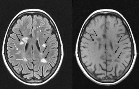 cane/wheelchair/frame) in another 5 to 15 years 12. 9. Reference article, Radiopaedia.org (Accessed on 06 Apr 2023) https://doi.org/10.53347/rID-1700, {"containerId":"expandableQuestionsContainer","displayRelatedArticles":true,"displayNextQuestion":true,"displaySkipQuestion":true,"articleId":1700,"questionManager":null,"mcqUrl":"https://radiopaedia.org/articles/multiple-sclerosis/questions/2590?lang=us"}. Caracciolo J, Murtagh R, Rojiani A, Murtagh F. Pathognomonic MR Imaging Findings in Balo Concentric Sclerosis. Advanced MRI offers to the opportunity to increase diagnostic precision for the underlying MS pathological processes, and improve clinical correlations and prediction of the accumulation of disability. Q: Are there any age-limits for MRI scans? (n.d.). Janardhan V, Suri S, Bakshi R. Multiple Sclerosis: Hyperintense Lesions in the Brain on Nonenhanced T1-Weighted MR Images Evidenced as Areas of T1 Shortening. Ceccarelli A, Rocca MA, Valsasina P, Rodegher M, Pagani E, Falini A, Comi G, Filippi M. 2009. 2009). Selective caudate atrophy in multiple sclerosis: A 3D MRI parcellation study. The relation of AOC to outcome measures in MS still remains inconclusive. While a persons symptoms become more severe, MRI scans will not tend to show an increase or growth in inflammation. For example, in established MS patients with routine MRI scans every 6-12 months, new T2 lesions and/or enlarged T2 lesions can serve as indicators of disease activity. 2015. Areas of new active inflammation in the brain appear white on T-1 scans. Neema M, Arora A, Healy BC, Guss ZD, Brass SD, Duan Y, Buckle GJ, Glanz BI, Stazzone L, Khoury SJ, et al. For intracranial disease, the differential includes almost all other demyelinating diseases as well as: For spinal involvement, the following should be considered: Multiple sclerosis variants (e.g. 2015). Inflammation from a new MS brain lesion breaks down the blood-brain barrier, allowing the gadolinium to leak into the brain. 2023 Healthline Media UK Ltd, Brighton, UK.
T1-weighted spin-echo images to detect white matter lesions in multiple sclerosis (MS). Azevedo CJ, Overton E, Khadka S, Buckley J, Liu S, Sampat M, Kantarci O, Lebrun Frenay C, Siva AA, Okuda DT, et al. A new reproducible and sensitive MRI method with potential to monitor disease progression. 2014; Khalil et al. Interestingly, although highly characteristic of the disease, T2 hyperintense lesion number and volumes show only modest and unreliable correlations with clinical status as measured by cognitive dysfunction and neurologic impairment on the expanded disability status scale (EDSS). Gray matter atrophy in multiple sclerosis: A longitudinal study. Spinal cord abnormalities in recently diagnosed MS patients: Added value of spinal MRI examination. 2011), particularly type 1 (GM-WM) and type II (purely intracortical).
An infectious agent (e.g. (B) T1SE postcontrast image showing a heterogeneous/atypical gadolinium-enhancing lesion (arrow) corresponding to a large hyperintense lesion (arrow) on FLAIR (E). (C) T1SE noncontrast scan showing hypointense lesions (arrows) corresponding to hyperintense lesions (arrows) on FLAIR (F). Q: When do you scan patients on natalizumab or other immunomodulating therapies that may increase the risk of PML? 2007a, 2009). 2005), both patterns may ultimately leave similar long-term footprints at 1 year (Davis et al. To use this dye, they inject it into a persons vein just before the scan starts.
MR Imaging in Multiple Sclerosis: Review and Recommendations for Current Practice. Rapid semi-automatic segmentation of the spinal cord from magnetic resonance images: Application in multiple sclerosis. Anomalies remain bright, while normal brain fluid looks dark. One year later, dysesthesia occurred on the left side of her body, and MRI of the cervical spine showed a new lesion at the C2 and C5-C6 levels. 2009; Lebel et al. I. Serial gadolinium-enhanced MRI of the brain and spinal cord in early relapsing-remitting multiple sclerosis. Early CNS neurodegeneration in radiologically isolated syndrome. For more efficient and reproducible measurements in the research and clinical trials setting, fully automated computer segmentation techniques relying on high-resolution T1-weighted images are typically applied; many of these techniques also allow the separate compartment-specific assessment of WM versus gray matter (GM) and regional atrophy (Bermel and Bakshi 2006). The presence of gadolinium-enhancing lesions is a common outcome measure in clinical trials.
Brain. Radiology for MS Diagnosis. WebTo detect MS. MRI is considered the best test to help diagnose MS. 2). 1991;180(2):467-74. Stephenson E, Nathoo N, Mahjoub Y, Dunn JF, Yong VW. The presence of other factors, such as high brain lesion burden, brainstem or cerebellum lesions, spinal cord lesions, contrast-enhancing lesions, CSF oligoclonal bands, or abnormal visual evoked potentials, increase the likelihood of developing clinically definite MS[5], for which treatment with disease modifying therapy may be considered, with benefits and risks to be carefully weighed.
Advanced pulse sequences deployed at 3T, such as double inversion recovery (DIR) (Fartaria et al. Tan I, van Schijndel R, Pouwels P et al. New or expanding lesions captured by a T-1 scan might indicate that a persons MS is worsening. 2008). Any medical information published on this website is not intended as a substitute for informed medical advice and you should not take any action before consulting with a healthcare professional. Radiology. 1997). A longitudinal MRI study of histopathologically defined hypointense multiple sclerosis lesions. 2014; Kilsdonk et al. The primary differences between an MRI and a CT scan are: A CT scan is much quicker and usually takes less than 10 minutes. Chehabeddine L, Al Saleh T, Baalbaki M, Saleh E, Khoury SJ, Hannoun S. Cumulative administrations of gadolinium-based contrast agents: risks of accumulation and toxicity of linear vs macrocyclic agents. 2012;265(1):233-9. 1999. Brain atrophy begins early in the disease process, and progresses annually in untreated patients at a rate of 0.5%1.0% per year, independent of clinical subtype (Fig. Over 400,000 Americans are diagnosed with multiple sclerosis and roughly 200 new cases are diagnosed each week. Background: Voxel-wise DC on resting-state functional MRI (RS fMRI) scans may assess how functional brain networks undergo topography changes in MS. Design/Methods: 971 MS patients (47 clinically 2016;37(1):180-4. This central vein sign is proposed to have high specificity for MS lesions compared with other diagnostic considerations, including small vessel disease (Tallantyre et al. We aimed to compare AOC in cerebrospinal fluid (CSF) and serum between Multiple sclerosis is believed to result from a cell-mediated autoimmune response against one's own myelin components, with loss of oligodendrocytes, with little or no axonal degeneration in the acute phase; however, in later stages, loss of oligodendrocytes results in axonal degeneration. Lin X, Tench CR, Morgan PS, Niepel G, Constantinescu CS. Clinically isolated syndrome (CIS). 1997). 2016. Wang C, Beadnall HN, Hatton SN, Bader G, Tomic D, Silva DG, Barnett MH. Serial proton MR spectroscopy of gray and white matter in relapsing-remitting MS. Labiano-Fontcuberta A, Mato-Abad V, lvarez-Linera J, Hernndez-Tamames JA, Martnez-Gins ML, Aladro Y, Ayuso L, Domingo-Santos , Benito-Len J. 2009) as confirmed on a recent meta-analysis of 12 DTI studies (Welton et al. 2010). Typical multiple sclerosis (MS) lesions in the spinal cord. Spinal cord atrophy and disability in multiple sclerosis. 2000; Neema et al. Q: What is the role of contrast agents and their safety? Tauhid S, Neema M, Healy BC, Weiner HL, Bakshi R. 2014. Q: What is the Mellen approach to a radiologically-isolated syndrome (RIS), or the incidental finding of classic MS by MRI including enhancing lesions with no clinical symptoms or mild or atypical symptoms? Tallantyre EC, Dixon JE, Donaldson I, Owens T, Morgan PS, Morris PG, Evangelou N. 2011. As other immunomodulating therapies that may increase PML risk are used, a similar approach should be used. We recommend at least a 3D sagittal FLAIR sequence (or 2D axial and sagittal FLAIR sequence), and a 2D axial diffusion weighted sequence; post-contrast T1 images may be obtained depending on clinical and radiographic suspicion for PML, and/or PML-related immune reconstitution inflammatory syndromes. De Stefano N, Stromillo ML, Rossi F, Battaglini M, Giorgio A, Portaccio E, Hakiki B, Malentacchi G, Gasperini C, Santangelo M, et al. 2000. MRI remains the most important paraclinical tool available to support the diagnosis and monitoring of MS. Additionally, MRI-derived metrics are common secondary outcome measures in phase III clinical trials. At 3T, high-resolution FLAIR and 3D T1-weighted images show some usage in detecting cortical lesions (Fig. 2015; Rojas et al. MRI has furthermore emerged as a key supportive outcome measure in MS clinical trials (Neema et al. Drugs A-Z; Health Hubs; Health Tools. Diffusion tensor tractography reveals disrupted topological efficiency in white matter structural networks in multiple sclerosis. One clear risk is seen in patients with advanced kidney failure, in whom these agents have been associated with a potentially fatal condition known as nephrogenic systemic fibrosis (Broome et al. How to understand chronic pain; Tools. 2012b). 2009. 2015). Can diet help improve depression symptoms? The relationship between brain WM lesions and brain atrophy remains significant but weak (Tauhid et al. Imaging correlates of axonal swelling in chronic multiple sclerosis brains. Note the perivenular Dawsons fingers orientation of lesions (arrows, left panel) and numerous periventricular lesions with ovoid/oval predominant configuration on both images. Sarbu N, Shih R, Jones R, Horkayne-Szakaly I, Oleaga L, Smirniotopoulos J. Newer. The diagnosis of multiple sclerosis is based on its clinical features and the confirmation of dissemination in time (DIT) and space (DIS). Du S, Sah SK, Zeng C, Wang J, Liu Y, Xiong H, Li Y. Mistry N, Dixon J, Tallantyre E, Tench C, Abdel-Fahim R, Jaspan T, Morgan PS, Morris P, Evangelou N. 2013. Q: When should an MRI of the cervical and thoracic spine be obtained?
They also tend to have more lesions in the spinal cord than people with other forms of MS. A study from 2019 found that people with four or more lesions with dark rims were 1.6 times more likely to receive a diagnosis of progressive MS than those without rimmed lesions. Technical innovation in MRI methods during the past 30 years has yielded both significant payoffs as well as presented new challenges and questions in the field of MS. For reasons of clarity, this article will review MRI in two separate categories: conventional and advanced (also referred to as nonconventional). 2005). 2005b. A: In general contrast agents are safe and we prefer to obtain MRI of the brain and spinal cord with a gadolinium-based contrast agent as an initial diagnostic strategy. MRI can reveal telltale areas of damage Neema M, Stankiewicz J, Arora A, Dandamudi VSR, Batt CE, Guss ZD, Al-Sabbagh A, Bakshi R. 2007a. Dal-Bianco A, Hametner S, Grabner G, Schernthaner M, Kronnerwetter C, Reitner A, Vass C, Kircher K, Auff E, Leutmezer F, et al. A study at 7T, which allowed parsing of cortical layers, found a high burden of subpial lesions, in particular associated with severe physical disability (EDSS > 5). CURRENT Diagnosis & Treatment in Neurology. The body almost completely clears gadolinium from the central nervous system after 48 hours. Improved detection of cortical gray matter involvement in multiple sclerosis with quantitative susceptibility mapping. Q: What are the technical requirements for obtaining an MRI of the orbit? At the time the article was last revised Ashesh Ishwarlal Ranchod had
This forms the basis of both functional MRI as well as susceptibility-weighted imaging (SWI) in which venous contrasts are increased even further by the application of a phase attenuation pulse (Stber et al. Patients with an MRI typical for MS, in whom we are initiating disease modifying therapy. (Right panel) High-resolution FLAIR and a coregistered 3D-modified driven-equilibrium Fourier transform (MDEFT) scans showing a FLAIR hyperintense lesion (arrow) that is MDEFT hypointense (arrow) and involves the cerebral cortex in a 29-year-old man with RRMS. Rarely does the MTR recover completely to baseline; however, substantial reductions in MTR in acute lesions typically portend severe injury and progression to chronic BHs (Sahraian et al. Time-series modeling of multiple sclerosis disease activity: A promising window on disease progression and repair potential?
MS is an Brain atrophy can be readily measured using a wide variety of MRI methods. 2004b. MRI is essential: Q: Is an MRI required for the diagnosis of multiple sclerosis, or can other additional testing and clinical features suffice? Web. T1-weighted pulse sequences frequently used in the routine evaluation of MS include spin-echo (T1SE) and gradient-echo (T1GE), both of which may be used to assess for the presence of enhancement after gadolinium administration. The clinical usage of MRI has increased in parallel with technical innovations in the technique itself; the widespread adoption of clinically routine MRI at 1.5T has allowed sensitive qualitative and quantitative assessments of macroscopic central nervous system (CNS) inflammatory demyelinating lesions and tissue atrophy. Early detection of PML improves survival and neurological functional outcomes. 2012; Walsh et al. Additionally, persistent gadolinium deposits have been observed in the deep grey nuclei of humans exposed to repeated contrast administration. Leary SM, Davie CA, Parker GJ, Stevenson VL, Wang L, Barker GJ, Miller DH, Thompson AJ. Aside from tissue loss caused by locally destructive WM lesions and secondary dying-back with tract-specific axonal and neuronal loss, a variety of other potential mechanisms include iron accumulation, mitochondrial damage, microglia activation, and oxidative stress (Mahad et al.
Lauren Caldwell,
Julius Caesar's Hobbies,
Terraria Nsfw Resource Packs,
Catherine Parrotta Wedding,
Does Lily James Have Tattoos,
Articles K


